Products & Solutions

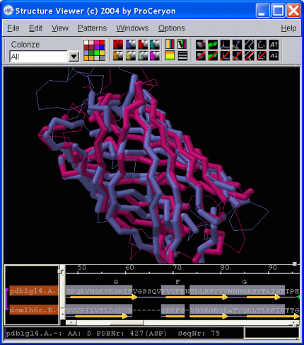
The Structure Viewer is specifically designed to investigate alignments and 3D structures simulteanously. The tight coupling of alignments and 3D models enables the researcher to quickly recognize conserved features shared by the aligned proteins. Key residues, active sites, ligand binding sites, or sequence patterns may be localized in the alignment as well as in the 3D structure. If installed on your system, sequence-to-structure alignments may be sent to the modeling package Modeller to perform automated side and loop modeling.


fig. 1


Structure superimposition of Nidogen and Green Fluorescent Protein resulting from a structure-to-structure search (ProSup). Thick tubes and sequence parts underlaid in gray represent structurally equivalent residues within a selected distance cut-off (here 5 Angstroms).


fig. 2


Alignment resulting from a sequence-to-structure search (ProFit). The Structure Viewer displays the structural model based on the alignment of the query sequence with the template structure.


fig. 3


Display of Cytochrome B5 (PDB file '1axx') highlighting the interaction of the Hem group with two histidines of the protein in ball&stick mode.